
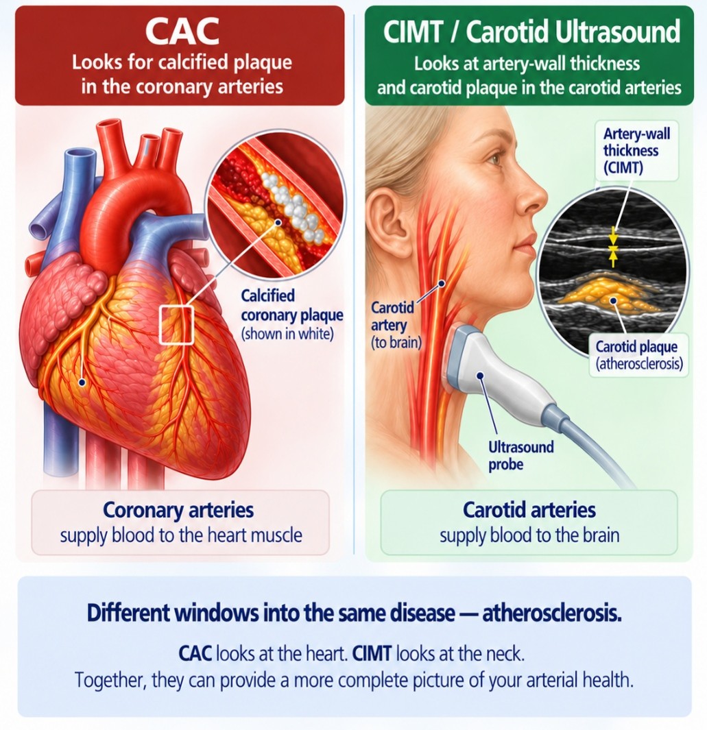
Two different windows into atherosclerosis—and how they can help personalize cardiovascular prevention
Most people learn about their cardiovascular risk through blood tests such as cholesterol, LDL, triglycerides, and blood sugar. More advanced testing may include ApoB, lipoprotein(a), insulin, and inflammatory or metabolic markers.
These tests are important because they help identify risk factors that may contribute to cardiovascular disease. But sometimes we want to answer a different question: Has atherosclerosis already started developing in my arteries?
Two noninvasive imaging approaches can help us look for evidence of atherosclerosis: a Coronary Artery Calcium (CAC) scan and carotid ultrasound, including carotid intima-media thickness (CIMT) and carotid plaque assessment.
The tests are not interchangeable. They examine different arteries and different features of atherosclerosis.
What Is Atherosclerosis?
Atherosclerosis is the gradual development of plaque within the walls of our arteries.
It usually develops over many years. Cholesterol-containing particles can enter and become trapped in the artery wall, where they can trigger inflammatory and immune responses. Other factors, including high blood pressure, abnormal blood sugar and insulin regulation, smoking, and genetics, can further influence the development and progression of atherosclerosis.
Over time, these processes can lead to the formation and growth of plaque within the artery wall. Plaque can also change in composition as it develops and may eventually become partially calcified.
Why does this matter? Because blood tests and imaging tests tell us different things about cardiovascular health.
Blood tests can help identify factors that may promote atherosclerosis. Imaging can help answer another question: Is there already visible evidence that atherosclerosis has developed?
The two imaging approaches discussed here provide different views of arterial health.
CoronaryArtery Calcium (CAC) Scan: Looking at the Heart
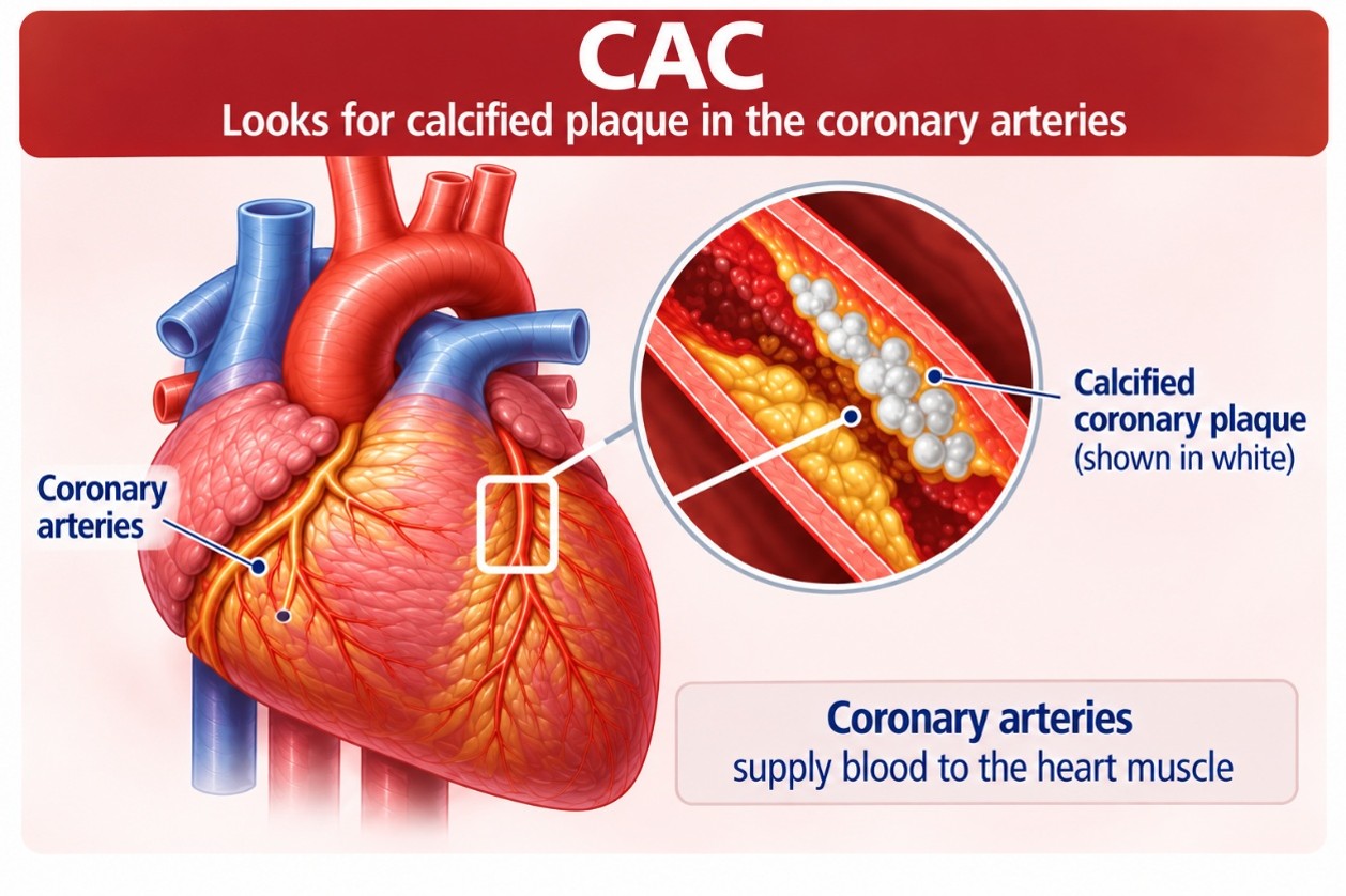
A coronary artery calcium scan is a specialized CT scan ofthe heart performed without contrast dye. It looks for calcium deposited withinatherosclerotic plaque in the coronary arteries, the arteries that supply bloodto the heart muscle.
The amount of coronary calcium detected is reported as a CAC score, commonly calculated using the Agatston scoring method. In simple terms, the higher the CAC score, the greater the amount of calcified coronary plaque detected. A score of zero means that the scan did not detect coronary artery calcium. Increasing scores indicate an increasing burden of calcified coronary atherosclerosis.
This matters because coronary calcium burden is stronglyassociated with future cardiovascular risk.
Why Is the CAC Score Useful?
One of the greatest strengths of CAC is risk prediction.
Consider two people of similar age with similar cholesterol levels and blood pressure. On paper, their calculated cardiovascular risk might appear similar.
But suppose
- Person A has CAC = 0
- Person B has CAC = 350
These individuals clearly do not have the same demonstrated burden of coronary atherosclerosis. That information can change the conversation about prevention.
CAC is particularly useful when a person's cardiovascular risk is uncertain and the patient and clinician are deciding how aggressively risk factors should be treated.
It can help answer questions such as
- Is there evidence that coronary atherosclerosis is already present?
- Does the person's estimated risk match what we can actually see in the coronary arteries?
- Should cholesterol-lowering treatment be considered or intensified?
- Should other cardiovascular risk factors be investigated more thoroughly?
- How aggressively should lifestyle and metabolic risk factors be addressed?
What Doesn't a Calcium Score Tell Us?
A coronary calcium scan does not detect all coronary plaque.
It is very good at detecting calcified plaque, but it does not adequately detect or characterize noncalcified plaque, sometimes informally called “soft plaque.”
Therefore, CAC = 0 does not necessarily mean, “I have absolutely no plaque.” It means that no coronary calcium was detected. It means: “No coronary calcium was detected.”
This distinction can be particularly important in younger individuals, in whom atherosclerosis may be developing before substantial calcification has occurred.
A CAC score also does not directly tell us whether a coronary artery has a significant blockage. If the clinical question is whether the coronary arteries are narrowed or whether noncalcified coronary plaque is present, another test—such as Coronary CT Angiography (CCTA)—may sometimes be more appropriate.
Does CAC = 0 Mean I Am Safe?
A CAC score of zero is generally very reassuring and is associated with substantially lower cardiovascular risk than measurable coronary calcium. But lower risk does not mean zero risk. A CAC score of zero still needs to be interpreted in the context of age, symptoms, cholesterol and ApoB, lipoprotein(a), diabetes, smoking, family history, and other cardiovascular risk factors.
CIMT and Carotid Ultrasound: Looking at the Neck
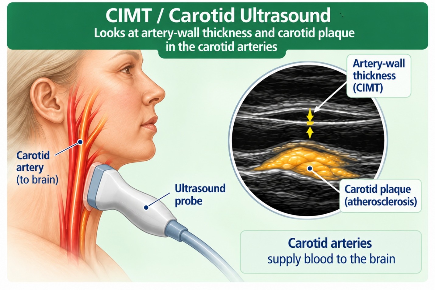
The carotid arteries run along each side of the neck and carry blood toward the brain. Because they are close to the skin, they can be examined noninvasively with ultrasound, which uses sound waves rather than ionizing radiation.
One measurement obtained during specialized carotid ultrasound is carotid intima-media thickness (CIMT). This measures the combined thickness of two layers of the carotid artery wall—the intima and media. The artery wall may become thicker with age and in response to cardiovascular risk factors.
However, increased CIMT and carotid plaque are not thesame thing.
CIMT measures the thickness of the artery wall, where as carotid plaque is a focal buildup within the arterial wall and provides more direct evidence that atherosclerosis is present.
For this reason, when carotid ultrasound is used to help assess cardiovascular risk, I am interested not only in the CIMT measurement, but also in whether carotid plaque is present and, if so, how much.
A comprehensive carotid ultrasound can provide information about arterial-wall thickness, the presence and location of plaque, overall plaque burden, and certain plaque characteristics.
An important advantage of ultrasound is that plaque does not have to contain calcium to be visible. Carotid ultrasound can therefore identify noncalcified as well as calcified plaque within the carotid arteries.
This helps explain why a person can have a CAC score of zero yet have plaque detected in the carotid arteries. The findings are not necessarily contradictory: the CAC scan looks for calcified plaque in the coronary arteries, while carotid ultrasound examines the carotid arteries and does not depend upon calcification to identify plaque.
The reverse can also occur—a person may have coronarycalcium while having relatively little carotid plaque.
Atherosclerosis does not necessarily develop at the samerate or to the same degree in every artery in the body.
Not all atherosclerotic plaque is the same.
Some plaques contain more lipid and inflammatory cells andrelatively little calcium. These are often referred to as noncalcified or“soft” plaques. Certain types of noncalcified plaque can be morebiologically active and more vulnerable to rupture. If a vulnerable plaqueruptures, a blood clot can form suddenly and potentially cause a heart attackor stroke.
As plaque evolves, portions of it may become calcified. Ingeneral, more densely calcified plaque tends to be more stable and less proneto rupture than lipid-rich, highly inflamed plaque.
However, this does not mean that coronary calcium isharmless. The presence of coronary calcium tells us that coronaryatherosclerosis is present, and a greater overall calcium burden is associatedwith greater cardiovascular risk.
This is why plaque composition and plaque burdentell us different things: the characteristics of an individual plaque mayinfluence its vulnerability, while the overall burden of atherosclerosis helpsus understand a person's broader cardiovascular risk.
CAC vs. CIMT/Carotid Ultrasound at a Glance
The simplest distinction:
- CAC asks: “How much calcified plaque can we see in the coronary arteries?”
- CIMT/carotid ultrasound asks: “What do the carotid artery walls look like, and is carotid plaque present?”
They are different windows into the same underlying disease process.
How Can These Tests Influence Treatment?
Neither test treats atherosclerosis. Their value is in helping us determine whether disease is already present and how aggressively cardiovascular risk should be evaluated and managed.
.jpg)
If imaging demonstrates atherosclerosis, the next question becomes: What may be driving the development of plaque in this individual?
The answer is not necessarily the same for everyone. Depending upon the individual, further evaluation may include:
- LDL cholesterol and ApoB
- lipoprotein(a), or Lp(a)
- triglycerides
- blood pressure
- glucose regulation and insulin resistance
- smoking exposure
- kidney function
- diet and body composition
- physical activity and cardiorespiratory fitness
- sleep quality and possible sleep apnea
- metabolic and inflammatory risk factors
- family history and genetics
Imaging findings may also influence decisions about cholesterol-lowering medications and the intensity of other preventive interventions.
For example, discovering substantial coronary calcium in someone previously thought to have only moderate cardiovascular risk may support a more intensive prevention strategy.
Conversely, a CAC score of zero may provide useful information when a treatment decision is uncertain—but it should never be interpreted in isolation from the rest of the person's cardiovascular risk profile.
The goal is not simply to lower cholesterol. The larger goal is to identify the factors driving atherosclerosis, reduce overall cardiovascular risk, and help prevent heart attack, stroke, and other vascular diseases.
Can These Tests Be Repeated to See Whether Treatment Is Working?
This requires some caution. It is tempting to assume that successful treatment should make a coronary calcium score decrease. That is not necessarily true.
Calcium represents only one component of atherosclerotic plaque. Effective lipid-lowering treatment can alter plaque composition and promote plaque stabilization, which may sometimes be accompanied by increased plaque calcification. Therefore, an increasing CAC score does not automatically mean that treatment has failed.
Similarly, small changes in CIMT over relatively short periods can reflect measurement variability rather than true progression or regression.
Neither test should therefore be treated like a cholesterol value that simply needs to decrease after treatment.
Whether either study should be repeated, and when, should be individualized based on the reason for testing and whether the result is likely to change clinical management.
FrequentlyAsked Questions
- Which test is better? Neither test is “better” for every purpose. CAC has the stronger evidence base for cardiovascular risk prediction and helping guide preventive treatment decisions. Carotid ultrasound provides different and potentially complementary information by examining the carotid artery wall and looking directly for plaque
- If my CAC score is zero, can I stop worrying about cholesterol? No. CAC = 0 is reassuring, but cardiovascular risk still depends on factors such as age, ApoB and LDL cholesterol, lipoprotein(a), blood pressure, diabetes, smoking, kidney disease, family history, and other individual risk factors.
- Can CIMT detect “soft plaque”? CIMT itself does not detect noncalcified (“soft”) plaque; it measures arterial-wall thickness. However, the carotid ultrasound examination can identify carotid plaque that does not need to be calcified to be visible. This is an important distinction because carotid ultrasound examines the carotid arteries, not the coronary arteries. It therefore cannot tell us whether noncalcified plaque is present in the coronary arteries.
- Which test involves radiation? A CAC scan uses CT and therefore involves a small amount of ionizing radiation. CIMT/carotid ultrasound uses sound waves and involves no ionizing radiation.
- Do either of these tests require contrast dye? No. A standard CAC scan does not require contrast dye, and neither does carotid ultrasound.
- If carotid plaque is present but my CAC is zero, does the carotid plaque matter? Yes. Visible carotid plaque is evidence of atherosclerosis and should be considered as part of the person's overall cardiovascular risk assessment. The findings are not contradictory because the tests examine different vascular territories and different aspects of plaque.
- Can either test tell me whether I have a dangerous blockage in my coronary arteries? Not reliably. CAC measures the burden of calcified coronary plaque. CIMT/carotid ultrasound examines arteries in the neck. If symptoms or other findings raise concern for significant coronary artery narrowing, additional cardiovascular evaluation may be necessary.
- Should everyone have both tests? No. Testing should have a purpose. For some people, CAC may provide the information needed. For others, carotid ultrasound may add useful information. In selected individuals, the two studies may be complementary. And in some patients, neither testis necessary because their medical history, symptoms, or existing cardiovascular disease already determine the appropriate evaluation and treatment.
The Bottom Line
Traditional cardiovascular risk factors remain important,but they primarily help us estimate cardiovascular risk. Imaging gives usanother perspective: Can we already see evidence of atherosclerosis?
A CAC scan looks for calcified atherosclerotic plaquein the coronary arteries of the heart.
CIMT/carotid ultrasound provides a radiation-freeview of carotid artery-wall thickness and carotid plaque.
Neither test tells the entire story. Used appropriately, and interpreted together with your medical history, family history, laboratory results, symptoms, and other cardiovascular risk factors, these studies can help make cardiovascular prevention more individualized.
The most useful question is therefore not simply, “Whatis my cholesterol?”
A better question may be: “Do I already have evidenceof atherosclerosis? What may be driving it, and what can I do now to helpprevent it from progressing?”
Which Test Is Appropriate for You?
Cardiovascular imaging should not be ordered simply because a test is available. The most useful test depends upon your age, symptoms, medical history, family history, laboratory findings, and existing cardiovascular risk factors.
If you are concerned about your cardiovascular risk, or if you have elevated cholesterol, ApoB or lipoprotein(a), high blood pressure, metabolic abnormalities, a strong family history of premature cardiovascular disease, or other risk factors, an individualized cardiovascular evaluation can help determine whether a CAC scan, CIMT/carotid ultrasound, additional laboratory testing, or another form of cardiovascular imaging would be most appropriate.
If you would like to better understand your cardiovascularrisk and determine which testing may be appropriate for you, please contact ouroffice to schedule an individualized evaluation.
This article is intended for educational purposes and does not replace individualized medical evaluation, diagnosis, or treatment.

